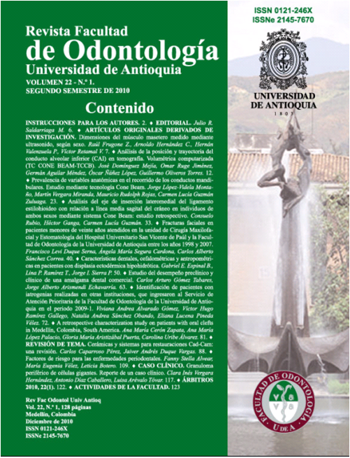
Dental, cephalometric and anthropometric characteristics in patients with hypohidrotic ectodermal dysplasia
DOI:
https://doi.org/10.17533/udea.rfo.2506Keywords:
Hypohisrotic ectodermal dysplasia, Anthropometric and cephalometric mesaurements, Anadontia, HypodontiaAbstract
Introduction: the objective of this study was to describe the facial and cephalometric characteristics of 16 patients with hypohidrotic ectodermal dysplasia (HED) being treated at the College of Dentistry of the University of Antioquia and to clinically and radiographically determine which teeth were present or absent; the age of the patients ranged between 5 and 19 years. Hypohidrotic ectodermal dysplasia is a genetic syndrome that mainly affects the embryonic ectodermal originated tissues, it is manifested as a triad which includes: hypotricosis, hypohidrosis and hypodontia; it is present in one of every one hundred thousand born alive. Intraorally, a delay in tooth eruption and conoid shaped teeth are observed, affecting both jaws. Other characteristics are: prominent front and, sunken nasal bridge, retrusion of the maxilla and, protrusion of the mandible. Methods: an univariate descriptive analysis was done using frequency tables, descriptive measurements, average, bar and pie graphs for the qualitative variables and frequency histograms for the quantitative variables. The statistical analysis was done with the SPSS data base, version 15.0. Results and conclusions: the decreased anthropometric measurements were: facial width (85.5%), cutaneous mandibular height for the upper lip (75%) and total upper lip height (56.3%). The increased measurements were: upper face height (81.3%), external inter canthal distance (68.8%) and forehead width (50%). At the skeletal level the cephalometric measurements showed Class III malocclusions with hypoplastic maxillas (62.5%), retrusion (81.3%), mandibles with adequate size and position and concave profiles (75%). The most commonly absent teeth were: upper and lower lateral incisors, upper first bicuspids and lower central incisors.
Downloads
Downloads
Published
How to Cite
Issue
Section
License
Copyright Notice
Copyright comprises moral and patrimonial rights.
1. Moral rights: are born at the moment of the creation of the work, without the need to register it. They belong to the author in a personal and unrelinquishable manner; also, they are imprescriptible, unalienable and non negotiable. Moral rights are the right to paternity of the work, the right to integrity of the work, the right to maintain the work unedited or to publish it under a pseudonym or anonymously, the right to modify the work, the right to repent and, the right to be mentioned, in accordance with the definitions established in article 40 of Intellectual property bylaws of the Universidad (RECTORAL RESOLUTION 21231 of 2005).
2. Patrimonial rights: they consist of the capacity of financially dispose and benefit from the work trough any mean. Also, the patrimonial rights are relinquishable, attachable, prescriptive, temporary and transmissible, and they are caused with the publication or divulgation of the work. To the effect of publication of articles in the journal Revista de la Facultad de Odontología, it is understood that Universidad de Antioquia is the owner of the patrimonial rights of the contents of the publication.
The content of the publications is the exclusive responsibility of the authors. Neither the printing press, nor the editors, nor the Editorial Board will be responsible for the use of the information contained in the articles.
I, we, the author(s), and through me (us), the Entity for which I, am (are) working, hereby transfer in a total and definitive manner and without any limitation, to the Revista Facultad de Odontología Universidad de Antioquia, the patrimonial rights corresponding to the article presented for physical and digital publication. I also declare that neither this article, nor part of it has been published in another journal.
Open Access Policy
The articles published in our Journal are fully open access, as we consider that providing the public with free access to research contributes to a greater global exchange of knowledge.
Creative Commons License
The Journal offers its content to third parties without any kind of economic compensation or embargo on the articles. Articles are published under the terms of a Creative Commons license, known as Attribution – NonCommercial – Share Alike (BY-NC-SA), which permits use, distribution and reproduction in any medium, provided that the original work is properly cited and that the new productions are licensed under the same conditions.
![]()
This work is licensed under a Creative Commons Attribution-NonCommercial-ShareAlike 4.0 International License.


