Archives
-
Revista Facultad de Odontología Universidad de Antioquia
Vol. 38 No. 1 (2026)Continuous publication
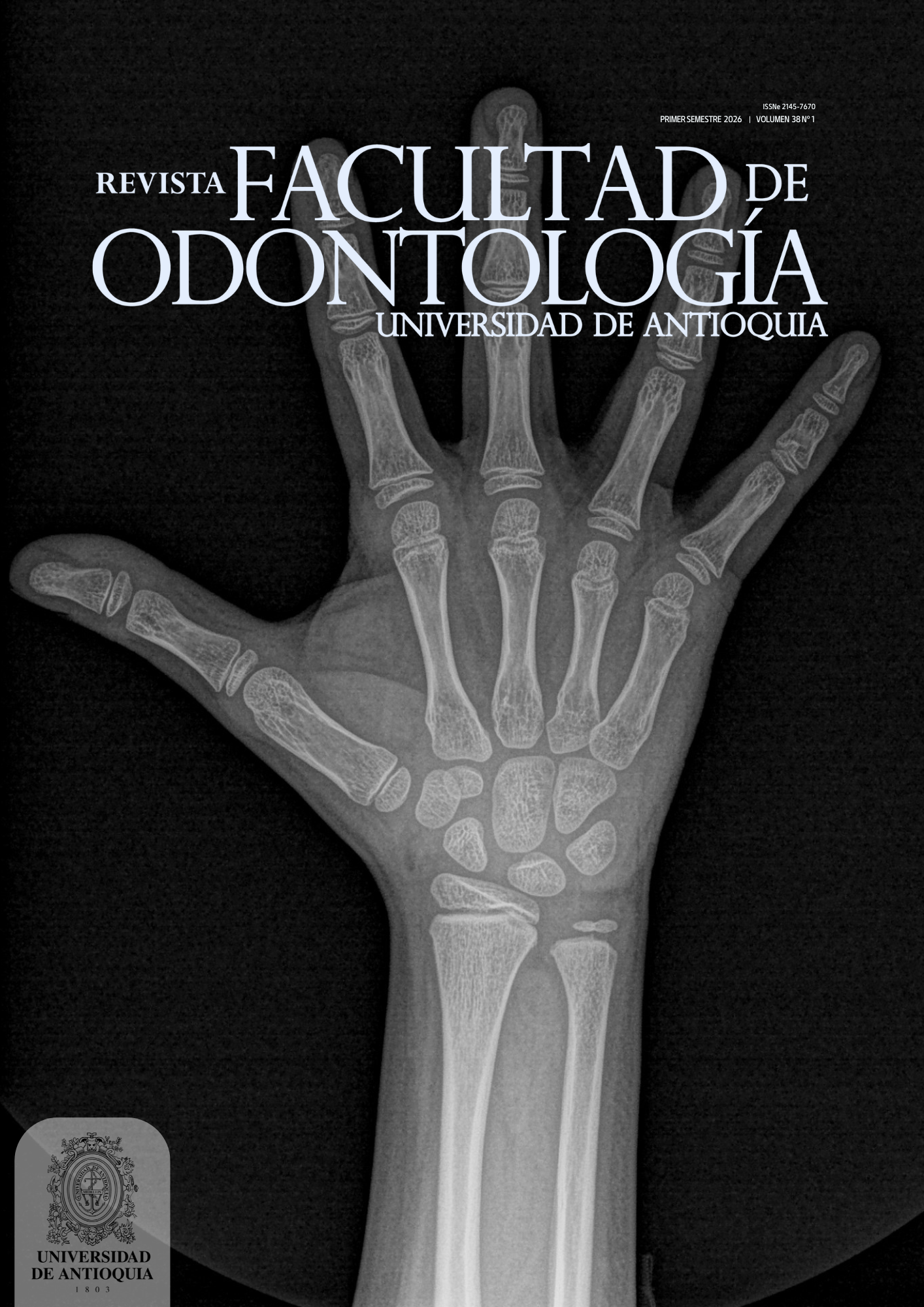
Cover: X-ray of a patient’s non-dominant hand showing different stages of bone maturation used to assess bone age. Various techniques have been applied, derived from different research studies, including the Fishman, Tanner–Whitehouse (TW3), and Greulich–Pyle (GP) methods.
X-ray taken at the Diagnostic Aid Center (DAC), Facultad de Odontología, Universidad de Antioquia.
Author: Pedro Jaramillo Vallejo
-

Revista Facultad de Odontología Universidad de Antioquia
Vol. 37 No. 2 (2025)Continuous publication
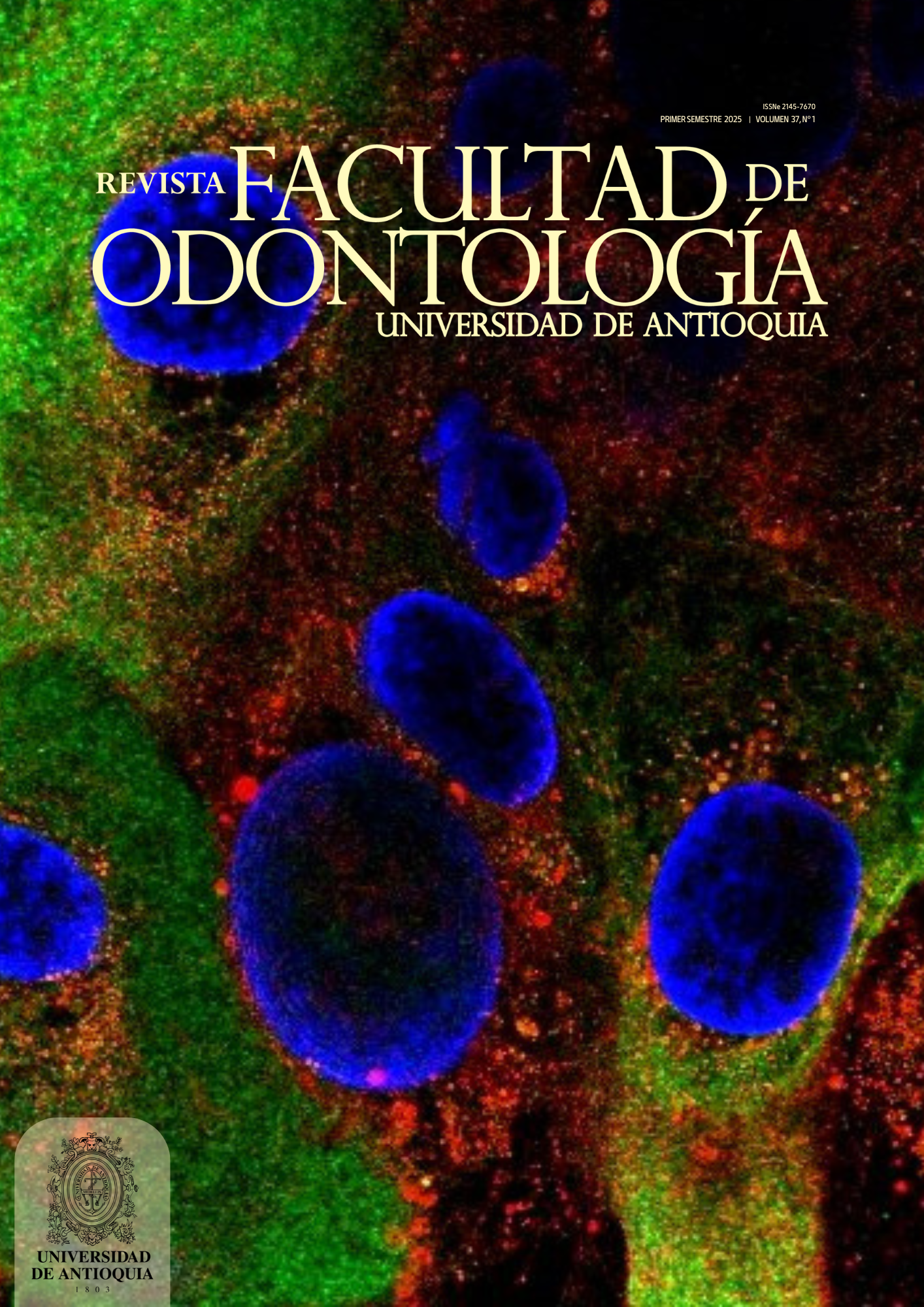
Cover: Confocal microscopy image of differentiated human oral keratinocytes (HOKs). HOKs serve as a valuable model for studying fundamental keratinocyte biology, as well as processes related to immortalization and malignant transformation. Immunocytochemistry was performed to detect ERP29 (green) and IRAK1 (red), while nuclei were stained with DAPI (blue). Images were captured and processed using a Leica SP8 Stellaris confocal microscope.
Author: Andrea Osorio, PhD(c) Facultad de Odontología, Universidad de Antioquia, BOA reseach group.
-
Revista Facultad de Odontología Universidad de Antioquia
Vol. 37 No. 1 (2025)Continuous publication
Cover: Confocal microscopy image of differentiated human oral keratinocytes (HOKs). HOKs serve as a valuable model for studying fundamental keratinocyte biology, as well as processes related to immortalization and malignant transformation. Immunocytochemistry was performed to detect IRF6 (green) and E-cadherin (red), while nuclei were stained with DAPI (blue). Images were captured and processed using a Leica SP8 Stellaris confocal microscope.
Author: Andrea Osorio, PhD(c) Facultad de Odontología, Universidad de Antioquia, BOA reseach group.
-

Revista Facultad de Odontología Universidad de Antioquia
Vol. 36 No. 2 (2024)July – December 2024
Cover: Untitled.
Author: Ana Isabel Correa-Orrego
-

Revista Facultad de Odontología Universidad de Antioquia
Vol. 36 No. 1 (2024)January – June 2024
Cover: Optical microscopy image of a chicken embryo (Gallus gallus) at embryonic day E5 in lateral view. This animal model is appropriate for the study of craniofacial development due to the great similarity with the one given in humans, its rapid formation, easy maintenance, manipulation and access in ovo.
Author: Mónica Parada, professor Facultad de Odontología, Universidad de Antioquia, BOA research group.
-

Revista Facultad de Odontología Universidad de Antioquia
Vol. 35 No. 2 (2023)July – December 2023
On the cover
Siproeta Stelenes
Author: Javier Enrique Botero Torres
-

Revista Facultad de Odontología Universidad de Antioquia
Vol. 35 No. 1 (2023)January – June 2023
On the cover
Ascalapha odorata
Author: Javier Enrique Botero Torres
-
Revista Facultad de Odontología Universidad de Antioquia
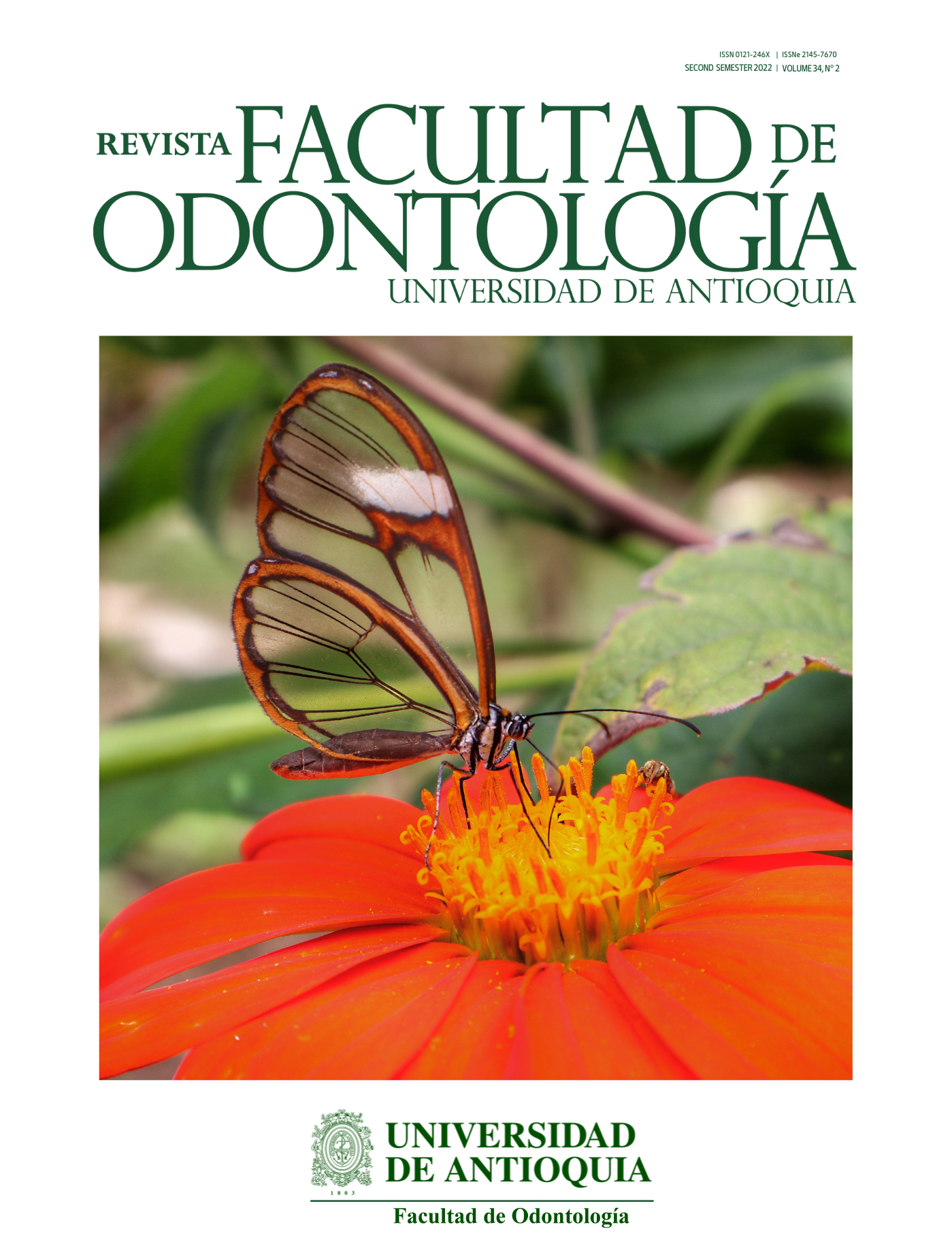
Vol. 34 No. 2 (2022)July – December 2022
On the cover
Dircenna adina
Author: Javier Enrique Botero Torres
-

Revista Facultad de Odontología Universidad de Antioquia
Vol. 34 No. 1 (2022)January – June 2022
On the cover
Marpesia corinna
Author: Javier Enrique Botero Torres
-
Revista Facultad de Odontología Universidad de Antioquia
Vol. 33 No. 2 (2021)July - December 2021
On the cover: Resurrexión
Author: Gonzalo de J. Moreno Moreno
Acrylic on wood
-
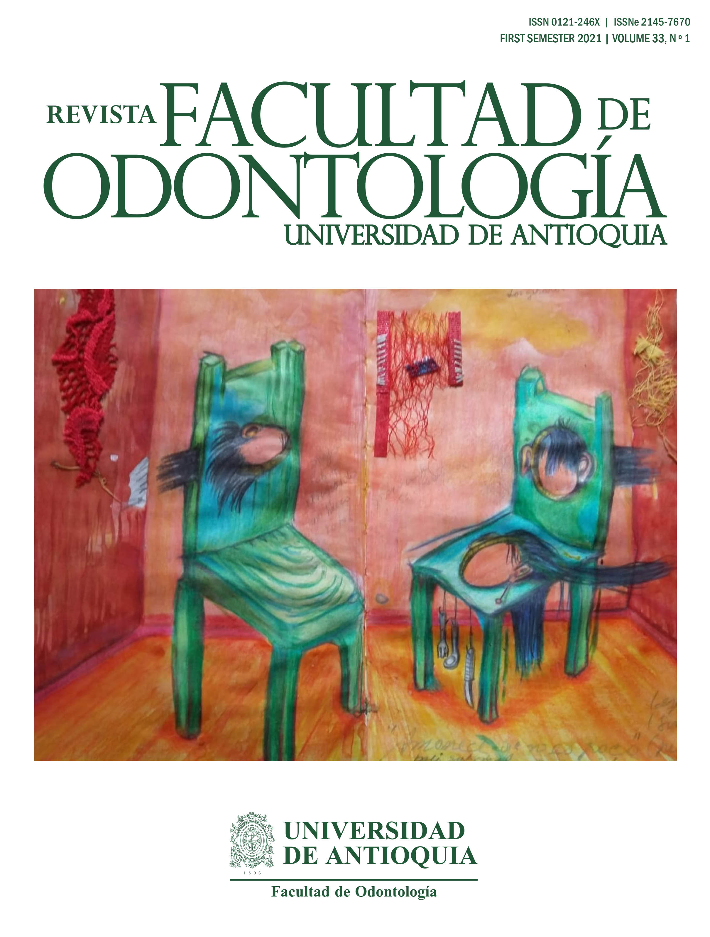
Revista Facultad de Odontología Universidad de Antioquia
Vol. 33 No. 1 (2021)January – June 2021
On the cover
Author: Elmer Restrepo
-
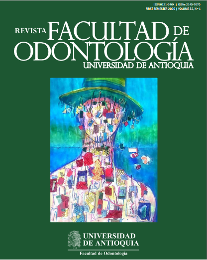
Revista Facultad de Odontología Universidad de Antioquia
Vol. 32 No. 2 (2020)July - December 2020
On the cover: Liria
Author: Elmer Restrepo, 2017
Watercolor and pencil
-
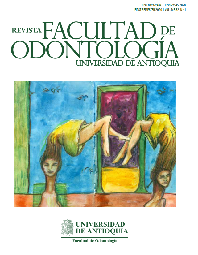
Revista Facultad de Odontología Universidad de Antioquia
Vol. 32 No. 1 (2020)January – June 2020
On the cover: Perro de Cristal
Author: Elmer Restrepo, 2020
Mixed technique
-
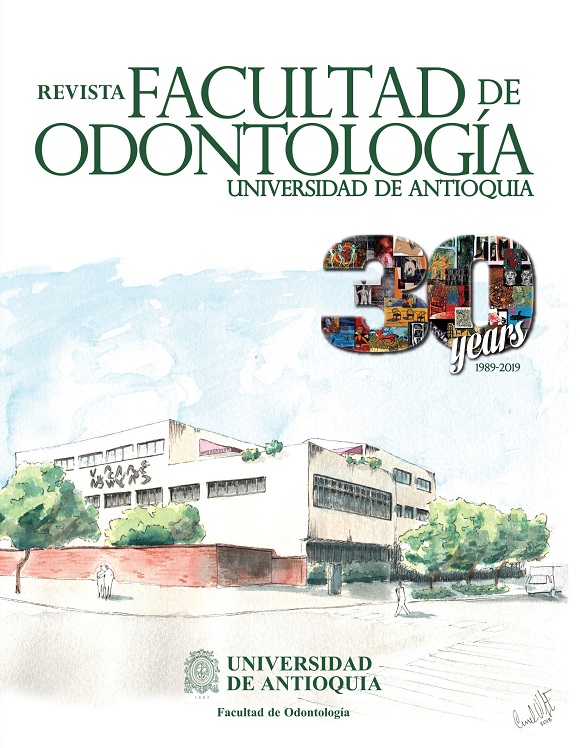
Revista Facultad de Odontología Universidad de Antioquia
Vol. 31 No. 1-2 (2019)July - December 2019
-

Revista Facultad de Odontología Universidad de Antioquia
Vol. 24 No. 1 (2012)Julio – Diciembre 2012