Nasal myiasis: report of a case and literature review
DOI:
https://doi.org/10.17533/udea.iatreia.v29n3a10Keywords:
Colombia, diabetes mellitus, diptera, ivermectin, myiasis, nasal avityAbstract
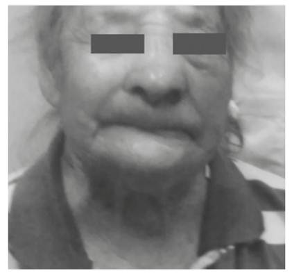
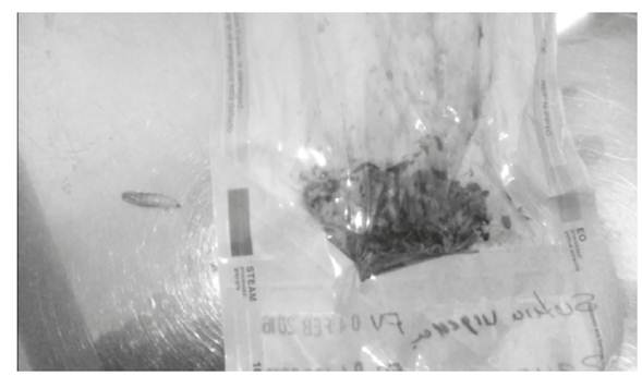
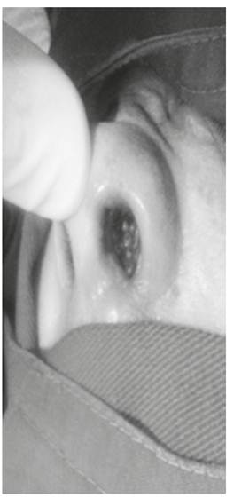
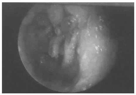
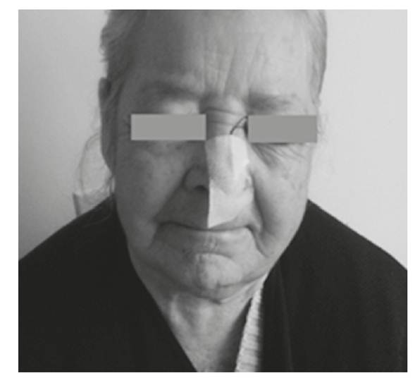
Myiasis is the infection of animal or human tissues or organs by larvae of Diptera. It may affect individuals of any age, but is more common in middle-aged and elderly patients. Nasal myiasis, an infection of the nasal and paranasal cavities by such larvae, is a common disease in tropical and developing countries. Reported cases of nasal myiasis have been caused by several different species, such as Lucilia sericata in Korea and Iran, Estro ovis in Algeria and France, Lucilia cuprina and Phaenicia sericata in Malaysia, Cochliomyia hominivorax in French Guiana, Drosophila melanogaster in Turkey, Eristalis tenax in Iran and Oestrus ovis in Israel. Signs and symptoms are related to the presence and movement of the larvae, and include foreign body sensation, bloody or muco-purulent nasal discharge. Prevention may be done with insect repellent. Treatment is based on antiparasitic drugs and techniques for removal of larvae, but may include the use of prophylactic topical or systemic antibiotics for possible secondary infections. We report a case of nasal and left maxillary sinus myiasis in an elderly woman, who responded favorably to treatment.
Downloads
References
(1.) Fierro-Arias L, Mercadillo-Pérez P, Sierra-Télles D, Puebla-Miranda M, Peniche-Castellanos A. Miasis furuncular en piel cabelluda: Reporte de un caso, presentación gráfica y revisión de la bibliografía. Dermatología CMQ. 2010;8(1):22-4.
(2.) Kim JS, Seo PW, Kim JW, Go JH, Jang SC, Lee HJ, et al. A nasal myiasis in a 76-year-old female in Korea. Korean J Parasitol. 2009 Dec;47(4):405-7. DOI 10.3347/kjp.2009.47.4.405.
(3.) Robbins K, Khachemoune A. Cutaneous myiasis: a review of the common types of myiasis. Int J Dermatol. 2010 Oct;49(10):1092-8. DOI 10.1111/j.1365-4632.2010.04577.x.
(4.) Moya J, Spelta MG, Gavazza S, Barbarulo AM, Fontana MI, Barerra M, et al. Miasis cutánea. Revisión sobre el tema y presentación de un caso de miasis forunculoide. Arch Argent Dermatol. 2007;57(5):217-22.
(5.) Cornet M, Florent M, Lefebvre A, Wertheimer C, Perez-Eid C, Bangs MJ, et al. Tracheopulmonary myiasis caused by a mature third-instar Cuterebra larva: case report and review. J Clin Microbiol. 2003 Dec;41(12):5810-2.
(6.) Manchini T, Fulgueiras P, Fente A. Miasis oral: a propósito de un caso. Odontoestomatología. 2009 May;11(12):38-43.
(7.) Reinoso-Quezada S, Alemán-Iñiguez JM. Rara miasis maxilar por Cochliomyia hominivorax. Reporte de caso, actualidad y entomología. Rev Esp Cir Oral Maxilofac. 2014. DOI 10.1016/j.maxilo.2014.04.005.
(8.) Thomas S, Nair P, Hegde K, Kulkarni A. Nasal myiasis with orbital and palatal complications. BMJ Case Rep. 2010 Dec;2010. DOI 10.1136/bcr.08.2010.3219.
(9.) Francesconi F, Lupi O. Myiasis. Clin Microbiol Rev. 2012 Jan;25(1):79-105. DOI 10.1128/CMR.00010-11.
(10.) González C, Salamanca J, Olano V, Pérez CE. Miasis cavitaria. Reporte de un caso. Rev Fac Med. 2008;16(1):95-8.
(11.) Aguilera A, Cid A, Regueiro BJ, Prieto JM, Noya M. Intestinal myiasis caused by Eristalis tenax. J Clin Microbiol. 1999 Sep;37(9):3082.
(12.) Chaccour C. Miasis forunculosa: Serie de 5 casos en indígenas de la etnia Pemón y revisión de la literatura. Dermatología Venezolana. 2005;43(4):8-15.
(13.) Africano FJ, Faccini-Martínez ÁA, Pérez CE, Espinal A, Bravo JS, Morales C. Pin-site myiasis caused by screwworm fly, Colombia. Emerg Infect Dis. 2015 May;21(5):905-6. DOI 10.3201/eid2105.141680.
(14.) Calvo LM, Suárez MM, Apolinario RM, Martín AM. Presencia de larvas en conducto auditivo externo y fosas nasales en paciente alcohólico. Enferm Infecc Microbiol Clin. 2005 May;23(5):323-4. DOI 10.1157/13074973.
(15.) Rosandiski Lyra M, Cruz Fonseca B, Sbragio Ganem N. Furuncular myiasis on glans penis. Am J Trop Med Hyg. 2014 Aug;91(2):217-8. DOI 10.4269/ajtmh.13-0688.
(16.) Salimi M, Edalat H, Jourabchi A, Oshaghi M. First Report of Human Nasal Myiasis Caused by Eristalis tenax in Iran (Diptera: Syrphidae). Iran J Arthropod Borne Dis. 2010;4(1):77-80.
(17.) Olea MS, Centeno N, Aybar CA, Ortega ES, Galante GB, Olea L, et al. First report of myiasis caused by Cochliomyia hominivorax (Diptera: Calliphoridae) in a diabetic foot ulcer patient in Argentina. Korean J Parasitol. 2014 Feb;52(1):89-92. DOI 10.3347/kjp.2014.52.1.89.
(18.) Babamahmoudi F, Rafinejhad J, Enayati A. Nasal myiasis due to Lucilia sericata (Meigen, 1826) from Iran: a case report. Trop Biomed. 2012 Mar;29(1):175-9.
(19.) Nazni WA, Jeffery J, Lee HL, Lailatul AM, Chew WK, Heo CC, et al. Nosocomial nasal myiasis in an intensive care unit. Malays J Pathol. 2011 Jun;33(1):53-6.
(20.) Mumcuoglu KY, Eliashar R. Nasal myiasis due to Oestrus ovis larvae in Israel. Isr Med Assoc J. 2011 Jun;13(6):379-80.
(21.) Sharma N, Malhotra D, Manjunatha BS, Kaur J. Oral Myiasis-A Case Report. Austin J Clin Case Rep. 2014;1(8):1039.
(22.) Visciarelli EC, García SH, Salomón C, Jofré C, Costamagna SR. Un caso de miasis humana por Cochliomyia hominivorax (Díptera: Calliphoridae) asociado a pediculosis en Mendoza, Argentina. Parasitol Latinoam. 2003 Jul;58(3-4):166-8. DOI 10.4067/S0717-77122003000300014.
(23.) Nene AS, Mishra A, Dhand P. Ocular myiasis caused by Chrysomya bezziana – a case report. Clin Ophthalmol. 2015 Mar;9:423-7. DOI 10.2147/OPTH.S79754.
(24.) Manfrim AM, Cury A, Demeneghi P, Jotz G, Roithmann R. Nasal Myiasis: Case Report and Literature Review. Int Arch Otorhinolaryngol. 2007 Jan-Mar;11(1):74-9.
(25.) Melendez HJ, Tamayo-Cáceres YR, Tello-Olarte YC, Vargas FO, Tarazona RA. Síndrome de dificultad respiratoria secundario a miasis sinusal y traqueopulmonar. Infectio. 2012 Jun;16(2):132-35. DOI 10.1016/S0123-9392(12)70068-1.
(26.) Ranga KR, Yadav SPS, Goyal A, Agrawal A. Endoscopic Management of Nasal Myiasis: A 10 Years Experience. AIJCR. 2013 Jun-Apr;6(1):58-60. DOI 10.5005/jp-journals-10013-1152.
(27.) De la Ossa N, Castro LE, Visbal L, Santos AM, Díaz E, Romero-Vivas CME. Miasis cutánea por Cochliomyia hominivorax (Coquerel) (Díptera: Calliphoridae) en el Hospital Universidad del Norte, Soledad, Atlántico. Biomédica. 2009 Jun-Mar;29(1):12-7.
(28.) Jimson S, Prakash CA, Balachandran C, Raman M. Oral myiasis: case report. Indian J Dent Res. 2013 Nov-Dec;24(6):750-2. DOI 10.4103/0970-9290.127626.
(29.) Osorio J, Moncada L, Molano A, Valderrama S, Gualtero S, Franco-Paredes C. Role of ivermectin in the treatment of severe orbital myiasis due to Cochliomyia hominivorax. Clin Infect Dis. 2006 Sep;43(6):e57-9.
(30.) Quesada-Lobo L, Troyo A, Calderón-Arguedas Ó. Primer reporte de miasis hospitalaria por Lucilia cuprina (Diptera: Calliphoridae) en Costa Rica. Biomédica. 2012;32(4):485-9. DOI 10.7705/biomedica.v32i4.690.
(31.) Sánchez-Sánchez R, Calderón-Arguedas O, Mora Brenes N, Troyo A. Miasis nosocomiales en América Latina y el Caribe: ¿Una realidad ignorada? Rev Panam Salud Pública. 2014;36(3):201-5.
(32.) Valderrama R, Arroyave M, Cadavid J, García P, Valencia P, Salazar C, et al. Prevalencia de miasis en el Hospital Universitario San Vicente De Paul (HUSVP), Medellín, Antioquia. Enero 1990–Marzo 2000. En: II Encuentro Nacional de Investigación en Enfermedades Infecciosas. Infectio [Internet]. 2000 [consultado 2016 May 3];4(1): [53]. Disponible en: http://revistainfectio.org/site/portals/0/ojs/index.php/infectio/article/view/384/398
(33.) Forero-Becerra EG. Miasis en salud pública y salud pública veterinaria. Una Salud. Revista Sapuvet de Salud Pública. 2011 Jul-Dic;2(2):95-132.
Published
How to Cite
Issue
Section
License
Copyright (c) 2016 Iatreia

This work is licensed under a Creative Commons Attribution-ShareAlike 4.0 International License.
Papers published in the journal are available for use under the Creative Commons license, specifically Attribution-NonCommercial-ShareAlike 4.0 International.
The papers must be unpublished and sent exclusively to the Journal Iatreia; the author uploading the contribution is required to submit two fully completed formats: article submission and authorship responsibility.


