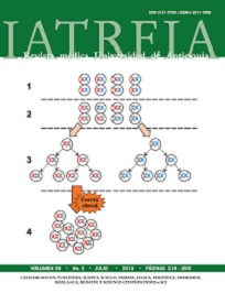
Determinación de la clonalidad en tejidos humanos
DOI:
https://doi.org/10.17533/udea.iatreia.v28n3a05Palabras clave:
cáncer, inactivación del cromosoma x, leucemia, linfoma, reordenamiento génicoResumen
Las proliferaciones malignas suelen ser clonales. La mayoría de las veces el potencial de una lesión se establece por medio del análisis clínico y el estudio anatomopatológico, pero algunos casos son de difícil diagnóstico. Por otra parte, existen situaciones en las que se producen clonas dominantes cuyo análisis es importante, tal como ocurre en enfermedades autoinmunes e inmunodeficiencias. Este artículo presenta de manera comprensible las técnicas principales para el estudio de la clonalidad, a saber: la evaluación de los reordenamientos génicos del receptor de antígeno y la evaluación del gen del receptor de antígeno humano.
Descargas
Citas
(1.) Greaves M, Maley CC. Clonal evolution in cancer. Nature. 2012 Jan;481(7381):306–13.
(2.) Swerdlow S, Campo E, Harris NL, Jaffe E., Pileri S., Stein H, et al. WHO Classification of Tumours of Haematopoietic and Lymphoid Tissues. 4th ed. Lyon: International Agency for Research on Cancer; 2008.
(3.) Rezuke WN, Abernathy EC, Tsongalis GJ. Molecular diagnosis of B- and T-cell lymphomas: fundamental principles and clinical applications. Clin Chem. 1997 Oct;43(10):1814–23.
(4.) Jaffe ES, Harris NL, Vardiman J, Elias Campo E, Arber DM. Hematopathology. St. Louis: Elsevier; 2010.
(5.) Goud KI, Dayakar S, Prasad SVSS, Rao KN, Shaik A, Vanjakshi S. Molecular diagnosis of lymphoblastic leukemia. J Cancer Res Ther. 2013 Jul;9(3):493–6.
(6.) Ståhlberg A, Aman P, Ridell B, Mostad P, Kubista M. Quantitative real-time PCR method for detection of B-lymphocyte monoclonality by comparison of kappa and lambda immunoglobulin light chain expression. Clin Chem. 2003 Jan;49(1):51–9.
(7.) van Dongen JJM, Langerak AW, Brüggemann M, Evans PAS, Hummel M, Lavender FL, et al. Design and standardization of PCR primers and protocols for detection of clonal immunoglobulin and T-cell receptor gene recombinations in suspect lymphoproliferations: report of the BIOMED-2 Concerted Action BMH4-CT98-3936. Leukemia. 2003 Dec;17(12):2257–317.
(8.) Gazzola A, Mannu C, Rossi M, Laginestra MA, Sapienza MR, Fuligni F, et al. The evolution of clonality testing in the diagnosis and monitoring of hematological malignancies. Ther Adv Hematol. 2014 Apr;5(2):35–47.
(9.) Perfetti V, Brunetti L, Biagi F, Ciccocioppo R, Bianchi PI, Corazza GR. TCRβ clonality improves diagnostic yield of TCRγ clonality in refractory celiac disease. J Clin Gastroenterol. 2012 Sep;46(8):675–9.
(10.) Sirsath NT, Channaviriappa LK, Nagendrappa LK, Dasappa L, Sathyanarayanan V, Setty GBK. Human immunodeficiency virus -associated lymphomas: a neglected domain. N Am J Med Sci. 2013 Jul;5(7):432–7.
(11.) Chen GL, Prchal JT. X-linked clonality testing: interpretation and limitations. Blood. 2007 Sep;110(5):1411–9.
(12.) Abbas AK, Lichtman AH, Pillai S, Igea Aznar JM. Inmunología celular y molecular. 6a ed. Barcelona: Elsevier; 2009.
(13.) Giallourakis CC, Franklin A, Guo C, Cheng H-L, Yoon HS, Gallagher M, et al. Elements between the IgH variable (V) and diversity (D) clusters influence antisense transcription and lineage-specific V(D)J recombination. Proc Natl Acad Sci U S A. 2010 Dec;107(51):22207–12.
(14.) Deriano L, Chaumeil J, Coussens M, Multani A, Chou Y, Alekseyenko AV, et al. The RAG2 C terminus suppresses genomic instability and lymphomagenesis. Nature. 2011 Mar;471(7336):119–23.
(15.) Knowles DM, Neri A, Pelicci PG, Burke JS, Wu A, Winberg CD, et al. Immunoglobulin and T-cell receptor beta-chain gene rearrangement analysis of Hodgkin’s disease: implications for lineage determination and differential diagnosis. Proc Natl Acad Sci U S A. 1986 Oct;83(20):7942–6.
(16.) Rombout PDM, Diss TC, Hodges E; Hummel M, van Dongen JJM, Langerak AW, Groenen PJTA. The Euro-Clonality website: information, education and support on clonality testing [internet]. J Hematopathol DOI 10.1007/s12308-011-0120-x Disponible en http://www.moleculardiagnostics.be/images/3SCM_Lange-rak_article1.pdf
(17.) Melotti CZ, Amary MFC, Sotto MN, Diss T, Sanches JA. Polymerase chain reaction-based clonality analysis of cutaneous B-cell lymphoproliferative processes. Clinics (Sao Paulo). 2010 Jan;65(1):53–60.
(18.) Harris S, Bruggemann M, Groenen PJTA, Schuuring E, Langerak AW, Hodges E. Clonality analysis in lympho-proliferative disease using the BIOMED-2 multiplex PCR protocols: experience from the EuroClonality group EQA scheme. J Hematop. 2012 Mar;5(1-2):91–8.
(19.) Van den Beemd R, Boor PP, van Lochem EG, Hop WC, Langerak AW, Wolvers-Tettero IL, et al. Flow cytometric analysis of the Vbeta repertoire in healthy controls. Cytometry. 2000 Aug;40(4):336–45.
(20.) Ribera J, Zamora L, Juncà J, Rodríguez I, Marcé S, Cabezón M, et al. Usefullness of IGH/TCR PCR studies in lymphoproliferative disorders with inconclusive clonality by flow cytometry. Cytometry B Clin Cytom. 2013 Jul 25; 21118(b):10.1002.
(21.) Kalina T, Flores-Montero J, van der Velden VHJ, Martin-Ayuso M, Böttcher S, Ritgen M, et al. EuroFlow standardization of flow cytometer instrument settings and immunophenotyping protocols. Leukemia. 2012 Sep;26(9):1986–2010.
(22.) Lima M, Almeida J, Santos AH, dos Anjos Teixeira M, Alguero MC, Queirós ML, et al. Immunophenotypic analysis of the TCR-Vbeta repertoire in 98 persistent expansions of CD3(+)/TCR-alphabeta(+) large granular lymphocytes: utility in assessing clonality and insights into the pathogenesis of the disease. Am J Pathol. 2001 Nov;159(5):1861–8.
(23.) Groenen PJTA, Langerak AW, van Dongen JJM, van Krieken JHJM. Pitfalls in TCR gene clonality testing: teaching cases. J Hematop. 2008 Sep;1(2):97–109.
(24.) Ortiz YM, Arias LF, Alvarez CM, García LF. Memory phenotype and polyfunctional T cells in kidney transplant patients. Transpl Immunol. 2013 Mar;28(2-3):127–37.
(25.) Vakiani E, Basso K, Klein U, Mansukhani MM, Narayan G, Smith PM, et al. Genetic and phenotypic analysis of B-cell post-transplant lymphoproliferative disorders provides insights into disease biology. Hematol Oncol. 2008 Dec;26(4):199–211.
(26.) Bonzheim I, Fröhlich F, Adam P, Colak S, Metzler G, Quintanilla-Martinez L, et al. A comparative analysis of protocols for detection of T cell clonality in formalin-fixed, paraffin-embedded tissue—implications for practical use. J Hematop. 2011 Dec 10;5(1-2):7–16.
(27.) Paireder S, Werner B, Bailer J, Werther W, Schmid E, Patzak B, et al. Comparison of protocols for DNA extraction from long-term preserved formalin fixed tissues. Anal Biochem. 2013 Aug;439(2):152–60.
(28.) Ghorbian S, Jahanzad I, Javadi GR, Sakhinia E. Evaluation diagnostic usefulness of immunoglobulin light chains (Igκ, Igλ) and incomplete IGH D-J clonal gene rearrangements in patients with B-cell non-Hodgkin lymphomas using BIOMED-2 protocol. Clin Transl Oncol. 2014 Nov;16(11):1006-11. DOI 10.1007/s12094-014-1188-4.
(29.) van Dongen JJM, Orfao A. EuroFlow: Resetting leukemia and lymphoma immunophenotyping. Basis for companion diagnostics and personalized medicine. Leukemia. 2012 Sep;26(9):1899–907.
(30.) Medeiros LJ, Carr J. Overview of the role of molecular methods in the diagnosis of malignant lymphomas. Arch Pathol Lab Med. 1999 Dec;123(12):1189–207.
(31.) Kosari F, Shishehbor F, Saffar H, Sadeghipour A. PCR-based clonality analysis in diffuse large B-cell lymphoma using BIOMED-2 primers of IgH (FR3) on formalin-fixed paraffin-embedded tissue. Arch Iran Med. 2013 Sep;16(9):526–9.
(32.) Hebeda KM, Van Altena MC, Rombout P, Van Krieken JHJM, Groenen PJTA. PCR clonality detection in Hodgkin lymphoma. J Hematop. 2009 Mar;2(1):34–41.
(33.) Beaufils N, Ben Lassoued A, Essaydi A, Dales J-P, Formisano-Tréziny C, Bonnet N, et al. Analysis of T-cell receptor-γ gene rearrangements using heteroduplex analysis by high-resolution microcapillary electrophoresis. Leuk Res. 2012 Sep;36(9):1119–23.
(34.) Boone E, Verhaaf B, Langerak AW. PCR-based analysis of rearranged immunoglobulin or T-cell receptor genes by GeneScan analysis or heteroduplex analysis for clonality assessment in lymphoma diagnostics. Methods Mol Biol. 2013 Jan;971:65–91.
(35.) Yang H, Xu C, Tang Y, Wan C, Liu W, Wang L. The significance of multiplex PCR/heteroduplex analysis-based TCR-γ gene rearrangement combined with laser-capture microdissection in the diagnosis of early mycosis fungoides. J Cutan Pathol. 2012 Mar;39(3):337–46.
(36.) Evans PAS, Pott C, Groenen PJTA, Salles G, Davi F, Berger F, et al. Significantly improved PCR-based clonality testing in B-cell malignancies by use of multiple immunoglobulin gene targets. Report of the BIOMED-2 Concerted Action BHM4-CT98-3936. Leukemia. 2007 Feb;21(2):207–14.
(37.) Langerak AW, Groenen PJTA, Brüggemann M, Beld-jord K, Bellan C, Bonello L, et al. EuroClonality/BIO-MED-2 guidelines for interpretation and reporting of Ig/TCR clonality testing in suspected lymphoproliferations. Leukemia. 2012 Oct;26(10):2159–71.
(38.) Bosaleh A, Denninghoff V, García A, Rescia C, Avagnina A, Elsner B. Rearreglos de genes de cadenas pesadas de las inmunoglobulinas en las gammapatías monoclonales. Medicina (B Aires). 2005;65(3):219–25.
(39.) Piña-Oviedo S, Fend F, Kramer M, Fournier F, Farca A, Ortiz-Hidalgo C. [Diagnosis of early gastric marginal zone lymphoma (MALT lymphoma) in endoscopic biopsies. Report of a case that demonstrates the utility of immunohistochemistry and the molecular analy-sis.]. Rev Gastroenterol México. 2008 Jan;73(3):172–6.
(40.) Novoa V, Núñez NA, Carballo OG, Lessa CF. Inmunofenotipos aberrantes en leucemias agudas en una población hospitalaria de Buenos Aires. Medicina (B Aires). 2013;73(1):9–16.
(41.) Quesnel B, Preudhomme C. [Residual disease: the hematologist’s point of view]. Bull Cancer. 2001 Jun;88(6):571–5.
(42.) Puig N, Sarasquete ME, Balanzategui A, Martínez J, Paiva B, García H, et al. Critical evaluation of ASO RQ-PCR for minimal residual disease evaluation in multiple myeloma. A comparative analysis with flow cytometry. Leukemia. 2014 Feb;28(2):391–7.
(43.) Cavé H, van der Werff ten Bosch J, Suciu S, Guidal C, Waterkeyn C, Otten J, et al. Clinical significance of minimal residual disease in childhood acute lymphoblastic leukemia. European Organization for Research and Treatment of Cancer--Childhood Leukemia Cooperative Group. N Engl J Med. 1998 Aug;339(9):591–8.
(44.) Garand R, Beldjord K, Cavé H, Fossat C, Arnoux I, Asnafi V, et al. Flow cytometry and IG/TCR quantitative PCR for minimal residual disease quantitation in acute lymphoblastic leukemia: a French multicenter prospective study on behalf of the FRALLE, EORTC and GRAALL. Leukemia. 2013 Feb;27(2):370–6.
(45.) Van Dongen JJ, Seriu T, Panzer-Grümayer ER, Biondi A, Pongers-Willemse MJ, Corral L, et al. Prognostic value of minimal residual disease in acute lym-phoblastic leukaemia in childhood. Lancet. 1998 Nov;352(9142):1731–8.
(46.) Brüggemann M, Schrauder A, Raff T, Pfeifer H, Dwor-zak M, Ottmann OG, et al. Standardized MRD quantification in European ALL trials: proceedings of the Second International Symposium on MRD assessment in Kiel, Germany, 18-20 September 2008. Leukemia. 2010 Mar;24(3):521–35.
(47.) Allen RC, Zoghbi HY, Moseley AB, Rosenblatt HM, Belmont JW. Methylation of HpaII and HhaI sites near the polymorphic CAG repeat in the human androgen-receptor gene correlates with X chromosome inactivation. Am J Hum Genet. 1992 Dec;51(6):1229–39.
(48.) Carrel L, Willard HF. X-inactivation profile reveals extensive variability in X-linked gene expression in females. Nature. 2005 Mar 17;434(7031):400–4.
(49.) Gong L, Ren K-X, Li Y-H, Liu X-Y, Zhang W-D, Yao L, et al. Determination of clonal status of pulmonary sclerosing hemangioma with X-chromosome inactivation mosaicism and polymorphism of phospho-glycerate kinase and androgen receptor genes. Med Oncol. 2011 Sep;28(3):913–8.
(50.) Mossner M, Nolte F, Hütter G, Reins J, Klaumünzer M, Nowak V et al. Skewed X-inactivation patterns in ageing healthy and myelodysplastic haematopoiesis determined by a pyrosequencing based transcrip-tional clonality assay. J Med Genet.2013 Feb; 50(2): 108-17.
Descargas
Publicado
Cómo citar
Número
Sección
Licencia
Derechos de autor 2015 Iatreia

Esta obra está bajo una licencia internacional Creative Commons Atribución-CompartirIgual 4.0.
Los artículos publicados en la revista están disponibles para ser utilizados bajo la licencia Creative Commons, específicamente son de Reconocimiento-NoComercial-CompartirIgual 4.0 Internacional.
Los trabajos enviados deben ser inéditos y suministrados exclusivamente a la Revista; se exige al autor que envía sus contribuciones presentar los formatos: presentación de artículo y responsabilidad de autoría completamente diligenciados.


