
Archives
-
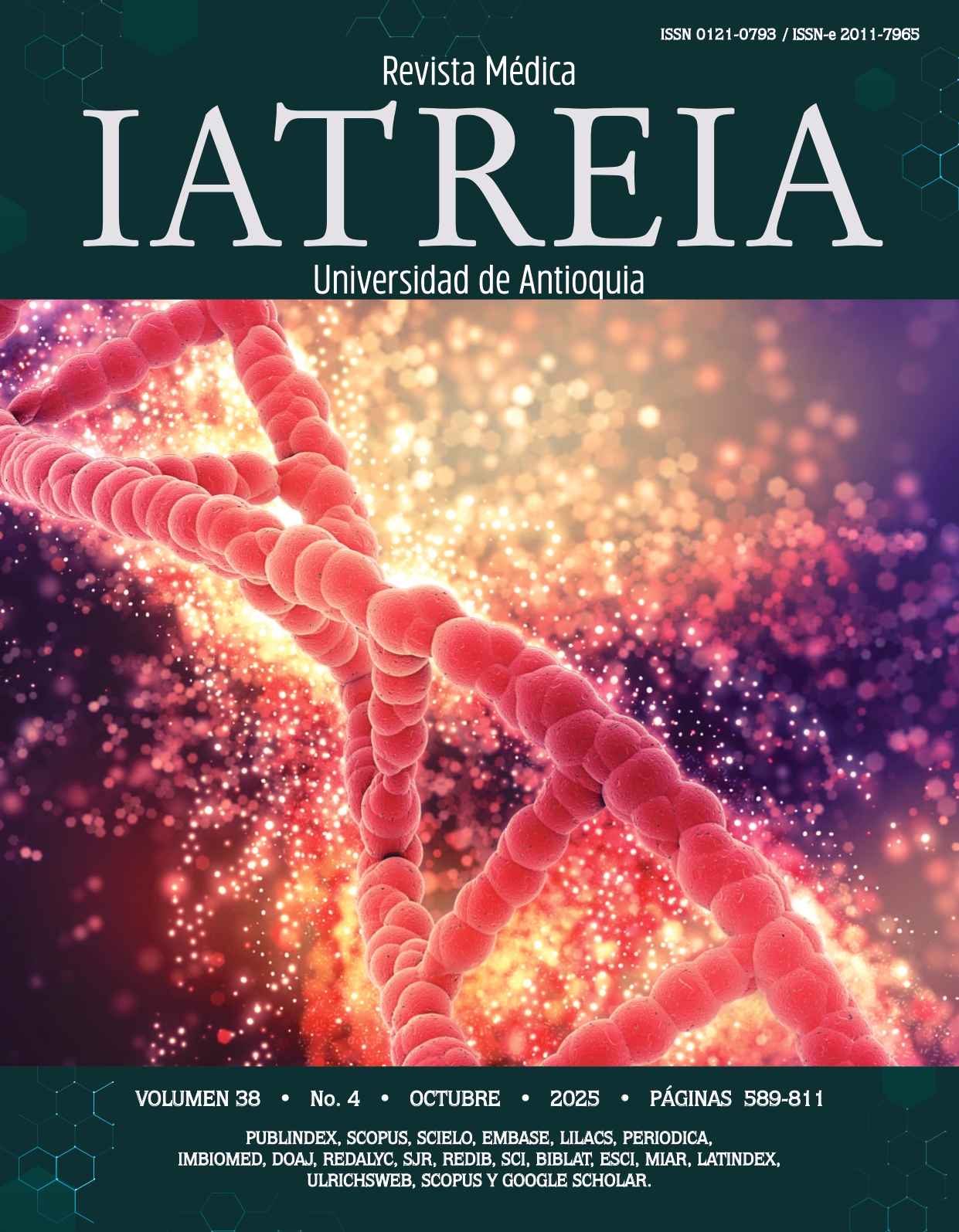
Vol. 38 No. 4 (2025)
Cover
The image accompanying the cover is inspired by the article “Epigenetic mechanisms associated with the pathogenesis of systemic lupus erythematosus: a review of the topic” published in this issue.Freepik. Hebra roja de ADN [Internet]. 2025 [cited 2025 Sep 9]. Available from:
https://bit.ly/45XIDbw -

Vol. 38 No. 3 (2025)
The illustration accompanying the third issue of volume 38 is inspired by the article “Biological Markers of Suicidal Behavior in Patients with Severe Mental Illness: a Narrative Review”, published in this issue.
Illustrator: Juan David Franco-Correa
-
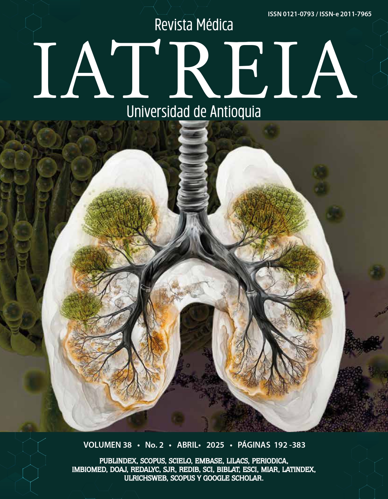
Vol. 38 No. 2 (2025)
The illustration accompanying the second issue of volume 38 is inspired by the article “Aspergillosis: Case Series from the Central Public Health Laboratory-Ministry of Health. Asunción, Paraguay. Period 2000 – 2019,” published in this edition.
Illustrator: Juan David Franco-Correa.
-
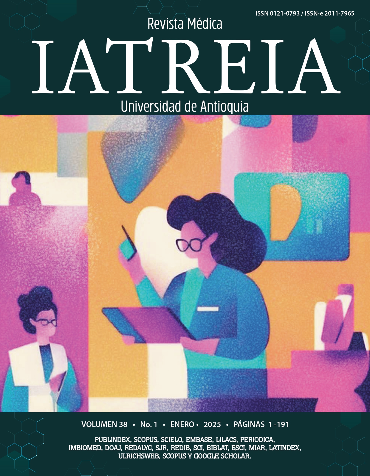
Vol. 38 No. 1 (2025)
The illustration that accompanies this first issue of volume 38 is inspired by the article “Characterization of executive functions and academic performance in medical students of the Corporación Universitaria Remington, 2021”, published in this edition.
Illustrator: Ana Laserna Lopera -

Vol. 37 No. 4 (2024)
This illustration is an allegory for two articles published in this issue: "Limbic Encephalitis: A Narrative Review of the Literature" and "Opioids in Pediatric and Neonatal ICU: A Proposal for Their Discontinuation." This metaphor is represented by the Papaver somniferum L. plant, from which opioids are derived and used for pain management. The article included in this issue proposes a strategy for discontinuing these medications, and the center of the plant depicts the brain affected by limbic encephalitis, in reference to the manuscript "Limbic Encephalitis: A Narrative Review of the Literature."
Design concept: Juan José Avilez
Illustrator: Ana Laserna Lopera
-

Vol. 37 No. 3 (2024)
This image was generated using Copilot artificial intelligence alluding to the article Prevalence and Factors Associated with Academic Burnout among Surgical Instrumentation Students in the Context of COVID-19 published in this issue.
-

Vol. 37 No. 2 (2024)
This image was generated using the Copilot artificial intelligence, referring to the article 'Incidence of Muscular Injuries in Professional Soccer Players: An Analysis before and during the COVID-19 Pandemic' published in this issue.
-

Vol. 37 No. 1 (2024)
HIV. Photo from the National Cancer Institute (@nci)
Taken from the free image bank Unsplash
Available at: https://bit.ly/3uWKo8x
-

Vol. 36 No. 4 (2023)
Photograph submitted to the Medical Photography and Illustration Contest of Iatreia Medical Journal.
Photograph Title: Membranous Glomerulonephritis
This photograph shows a glomerulus from a patient with membranous glomerulonephritis, displaying the characteristic spikes or projections of the basement membrane, stained with silver methenamine (original magnification 40X).
Morphologically, immune complexes deposit in the subepithelial capillary walls (outer face of the basement membrane), prompting a response from the membrane to try to encircle or surround them. In light microscopy, with silver methenamine staining, perpendicular projections can be observed on the basement membrane, consisting of material similar to it, known as spikes. These projections progressively extend and encircle or envelop the immune complexes. In tangential sections of the membrane, they appear as spaces or holes corresponding to the immune complex (an apparently empty space that does not stain with silver methenamine), completely surrounded by the basement membrane. These immune complexes, in some cases, can also be visualized by light microscopy with trichrome staining and, more specifically, by immunofluorescence.
Authors: Jeanneth Echeverri-Villegas, Julián Rondón-Carvajal
-

Vol. 36 No. 3 (2023)
Winners of the Iatreia Medical Journal's First Photography and Medical Illustration Contest
Title of the photograph: Spontaneous Pneumoperitoneum Secondary to Intestinal Cystic Pneumatosis
Description: A 40-year-old woman with a history of systemic lupus erythematosus (SLE) for a year and a half, presenting to the emergency department with visceral abdominal pain of two weeks' duration. She underwent a contrast-enhanced abdominal computed tomography (CT) scan, which revealed gas accumulation in the greater omentum and subserosal layer of the small intestine and colon, as well as secondary pneumoperitoneum in the coronal plane (Figure A). Due to the worsening of her clinical condition, diagnostic and therapeutic laparoscopy was performed, documenting multiple thin-walled air-filled cysts (cystic pneumatosis) in the colon and small intestine, without perforation of hollow viscera (Figure B). Finally, conservative medical management with parenteral steroid pulses was chosen, leading to an acceptable medical response over time, without additional surgical interventions.
Authors: Milena Alcázar-Paternina, Tatiana Suárez-Poveda, Vanessa García-Gómez, Julián Rondón-Carvajal
-

Vol. 36 No. 2 (2023)
Winners of the Iatreia Medical Journal's Photography and Illustration Contest
Photograph Title: "Diafanización" (Diafanization)
Description: A preserved fish and snake using diafanization, a technique that involves depigmenting soft tissues and pigmentation of mineralized tissues to visualize the bony and cartilaginous components of the organism being studied. This and other preserved animals can be found in the morphology building of the Faculty of Medicine at the University of Antioquia.
Author: Nicolás Sierra Paz
-

Vol. 36 No. 1 (2023)
Ganadores del I Concurso de Fotografía e Ilustración Médica Revista Iatreia
Título de la fotografía: Morfogénesis del virus de la viruela de mono
Descripción: composición que muestra las etapas de formación del virus de la viruela del mono (Monkeypox virus) en células de riñón de mono (VERO-E6). Las células infectadas fueron fijadas con glutaraldehído al 2,5 % y tetróxido de osmio al 1,2 %, se incluyeron en resina epóxica y se hicieron cortes de 60 a 90 nanómetros; los cortes se contrastaron con acetato de uranilo y citrato de plomo y se visualizaron en un microscopio electrónico de transmisión con un aumento de 1.000.000 X. A. Dos semilunas que constituyen la etapa inicial de la formación del virus. B. Viriones inmaduros (óvalos claros) y algunos en un estado más avanzado de formación (óvalos oscuros). C. Varios viriones maduros cerca a la membrana celular y un virión en el momento de salir de la célula. D. Virión maduro fuera de la célula; en este se pueden observar algunos detalles como la membrana de superficie (flecha corta), el core o nucleoide (flecha larga) y los cuerpos laterales (cabezas de flecha).
Autores: Francisco J. Díaz, Wbeimar Aguilar, María Isabel Zapata, María Teresa Rugeles, Grupo Inmunovirología, Universidad de Antioquia.
-
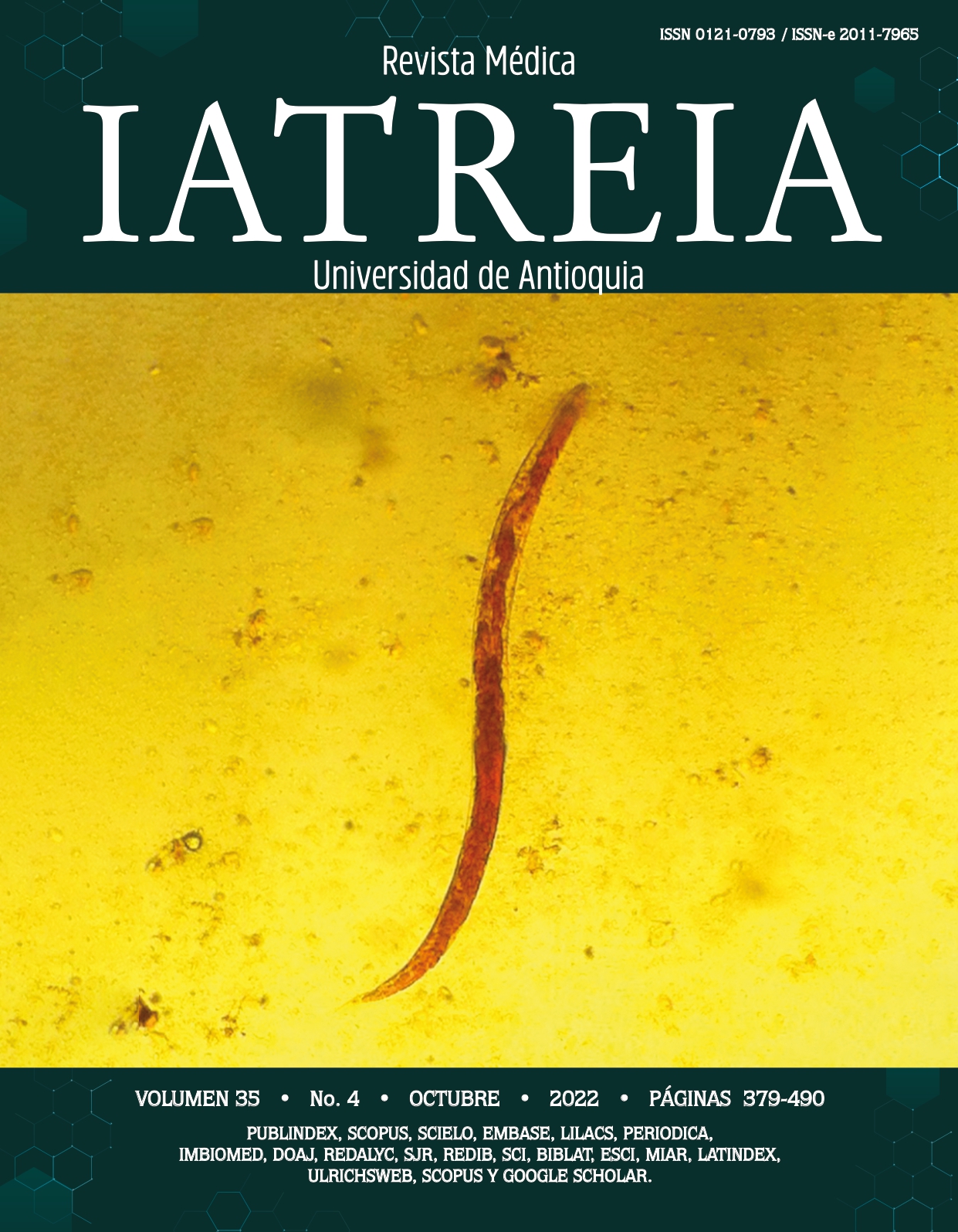
Vol. 35 No. 4 (2022)
Blueiridium. Strongyloides species [Internet]. [Consultado 2022 sept 29]. Disponible en: https://bit.ly/3CkG58t
-
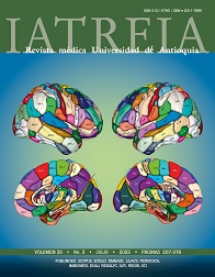
Vol. 35 No. 3 (2022)
Carátula: Esquema de parcelación del cerebro humano en el Brainnetome Atlas. Tomado de: Fan L,et al. DOI 10.1093/cercor/bhw157
-

Vol. 35 No. 2 (2022)
Cover: v2osk. Unsplash [internet]. [Consultado 2022 feb 15]. Disponible en: https://bit.ly/3oPYa7j
-
Vol. 35 No. 1 (2022)
Cover: Mac231. Book-Encyclopedia [internet]. [Accessed 2021 Dec 6]. Available from: https://acortar.link/cWq2K0
-
Vol. 34 No. 4 (2021)
Cover page: Jplenio. Virus [internet]. [Accessed 2021 Sep 10]. Available from: https://acortar.link/M3WODQ
-
Vol. 34 No. 3 (2021)
Cover: Inverted classroom in the virtual era (Dr. Diego Orlando Sierra, Professor Department of Surgery,
Universidad de la Sabana, Colombia).


1-25 of 169
Next


